

Virtual Surgical Planning for
Orthognathic Surgery:
Precise Bite Splint
The Precise Team’s expertise in Orthognathic Virtual Surgical Planning (VSP) assists in accurately visualising the full treatment plan and communicating it to the patient using the latest technology available.
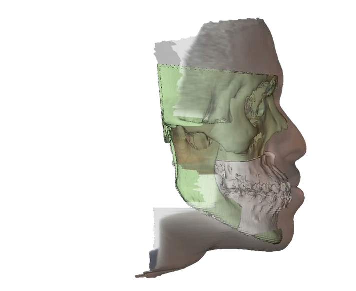
Analysis of the 3D Bony movements and the predicted soft-tissue changes are added to conventional planning tools allowing all parties involved to effectively visualize the treatment plan.
VSP allows planning of simple as well as complex 3D movements which are difficult to achieve using traditional methods alone. Digital technology removes manual processes leading to greater accuracy.
Intra-oral Scan
Virtual Surgical Planning
3D Printed Occlusal Splint
PROCEDURES
LeFort I
LeFort II
LeFort III
Segmental LeFort I
Bilateral and Unilateral Sagittal Split Osteotomy
Genioplasty
Multi-Jaw surgery with asymmetry
Orthognathic surgical planning with stock TMJ
BENEFITS
3D printed intermediate and final splints
Analyse anatomical discrepancies in three dimensions
Visualisation of the airway
Soft tissue simulation
Analyse how the occlusal movement will affect the surrounding bony anatomy and soft tissue
Compare multiple treatment plans for the same patient
Collaboration between the Surgeon and the Orthodontist
PRODUCTS
A 'precise' solution that is patient matched using the patients’ CT scan data.
Full treatment plan report
A 3D video of the
final bony and soft tissue movements
Soft tissue prediction
Intermediate splint
Final splint (optional)
ORTHOGNATHIC PLANNING INCLUDES:


Intermediate
and
Final Splints

Osteotomy
and Overlap Measurements

Soft Tissue
Simulation
If you would like an 'optional extra', let’s talk about your plans.
TIMELINE (9 DAYS)
DESIGN
DAY


Receive Scans,
Photos and
Stone Models
(2 Days)


Data Import and
preliminary
design


Surgeon Meeting
&
Approval*
Final images and
Case Report
3D
Printing
(2 Days)
Delivery

Bite splint
verification


*Timeline may be extended due to design changes